Posted October 2, 2013
Using nanoparticles to detect heart attack, stroke risk
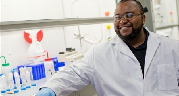
Joseph Labolito
Omar Fisher, assistant professor of bioengineering, is working to develop a nanotechnology that will help doctors determine the location and severity of plaques in artery walls before they rupture.
A Temple bioengineer is collaborating on an NIH-funded project to develop a nanotechnology that will help doctors determine the location and severity of plaques in artery walls before they rupture.
Atherosclerosis, or hardening of the arteries, occurs when fat, cholesterol, calcium and other substances form such plaques. The disease is a major cause of heart attacks and strokes when these plaques rupture and form clots in the blood.
Omar Z. Fisher, assistant professor of bioengineering in Temple’s College of Engineering, has developed a method for linking polyphenols, which are very strong antioxidants, to polymers that can self-assemble into nanoparticles.
Fisher said the project will focus on using these polymers to encapsulate superparamagnetic iron oxide particles (SPIOs), a nano-scale MRI contrasting agent used by his collaborator, Amber Doiron, assistant professor of bioengineering at Binghamton University.
The antioxidant nanoparticles containing the contrasting agent would travel through the blood vessels until they reach atherosclerotic plaque, enabling doctors to diagnose the severity of plaques before they rupture, said Fisher.
“The worse the plaque is, the more likely it is to rupture and give you a clot,” he said. “Because these polymers are made from antioxidants, they are sensitive to oxidative stress, which is more prevalent in more severe plaques.”
Once the oxidative stress destroys the polymers, the MRI contrast agents will be released inside the plaques.
Fisher said doctors would likely prescribe an MRI when a patient was showing some signs of cardiac distress such as fatigue or chest pains.
“During the MRI, the degree of contrast would be visible and indicate to doctors not only that the plaques were there, but the severity of the plaques and how likely they would be to rupture,” he said.
The project is supported through a two-year, $418,000 grant which Fisher and Doiron recently received from the National Institute for Biomedical Imaging and Bioengineering.