Posted February 16, 2010
Lasers could speed cancer testing: Photonics center explores new technology that might enable real-time diagnosis
 |
|
| Joseph V. Labolito / Temple University | |
|
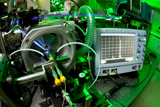
A doctor takes a tissue biopsy from a patient and sends it to a lab to be analyzed for cancer, and it can takes hours, days or, in some cases, even a week to get the results. Now a new analysis technology developed by researchers in Temple’s Center for Advanced Photonics Research could possibly tell whether tissue is cancerous or normal in a matter of seconds. “If you get cancer, the diseased tissue changes chemical composition,” said Robert Levis, chair of chemistry and director of CAPR. “If we can measure the tissue’s chemical composition, we can tell if it’s healthy tissue or abnormal.” Levis and his group have pioneered a method in which an ultra-fast laser is used to vaporize materials, molecules or tissues. Once in gas form, an electro-spray mass spectrometer weighs the materials’ bio-molecules. Vaporizing a sample on a plate with the femtosecond (one quadrillionth of a second) laser releases the molecules into the air, regardless of size or vapor pressure. The molecules are engulfed by the electro-spray, much like a hose spray can engulf a flower petal. The electrospray is a water methanol solution ejected from a hypodermic needle by a high voltage, and this ultimately places a charge on the molecules. A small capillary tube opposite the needle charged at -5,000 volts pulls the tiny positively charged droplets into a vacuum and then into the mass spectrometer. “It’s basically the same way they paint cars,” said Levis. “The droplets come out of the needle, they get charged and then they’re drawn to the five kilo-volts, making a beautiful even coating when you paint or a soft ionization method for mass spectrometry." “In our experiment, the charged molecule is flung down an evacuated tube, which is about a meter long, and we count the amount of time required to strike a charge detector,” said Levis, a pioneer in strong field chemistry. “The transit time reveals how massive these molecules are, and what’s really amazing is, we can analyze molecules as big as Mother Nature makes them.” So far, Levis’ group has examined biological molecules such as vitamin B12 and oxycodone before moving to leafs and plants as well as complex biological mixtures like blood. Graduate students John Brady and Elizabeth Judge directed the laser to different parts of an impatiens plant and saw different mass-spectral signatures for the stem, leaves and petals. “We even put a spot of blood down on the slide, vaporized it with the laser and the electro-spray and amazingly, we were able to weigh all of the hemoglobins in the mass spectrometer,” said Levis. “So we can differentiate tissue types, so now, I think we will be able to use this method to tell the difference between cancer tissue and normal tissue. If we can build a library of tissue signatures, we will be able to tell quickly exactly what certain type tissue is because our mass spectra are measured in real-time.” The researchers have published their initial findings in the journal, Rapid Communications in Mass Spectrometry. Levis said they are now looking to collaborate with researchers on liver disease and lung disease. |