Microscopy acquisitions bring scientific research into focus at Temple
The need for researchers to see and understand materials at increasingly high resolution has become greater and greater. Now more help is on the way to Temple researchers thanks to a recent $475,000 U.S. Department of Defense grant to purchase a scanning electron microscope.
Parsaoran Hutapea, associate professor of mechanical engineering, received the Defense University Research Instrumentation Program grant through the Office of Naval Research. Xiaoxing Xi, Laura H. Carnell Professor of Physics; Svetlana Neretina, assistant professor of mechanical engineering; and Nicholas Davatzes, assistant professor of earth and environmental science, are co-principal investigators on the grant.
The Defense University Research Instrumentation Program supports the DoD’s investment of more than $2 billion each year in basic, applied and advanced research at universities. Hutapea, Xi and Davatzes all receive research funding from the DoD.
“With the rise of nanotechnology-related research taking place on campus, there is a growing need for this type of microscope,” said Hutapea. “This microscope will allow us to produce realistic three-dimensional images of nano-scale specimens.”
Hutapea said that the new microscope will be used by researchers in mechanics, physics, materials engineering, earth and environmental science, biomedical engineering, medicine and chemistry. It will be installed in the College of Engineering and should be in operation no later than next spring.
This is the third major piece of microscopy equipment to be acquired by Temple researchers in the past three years.
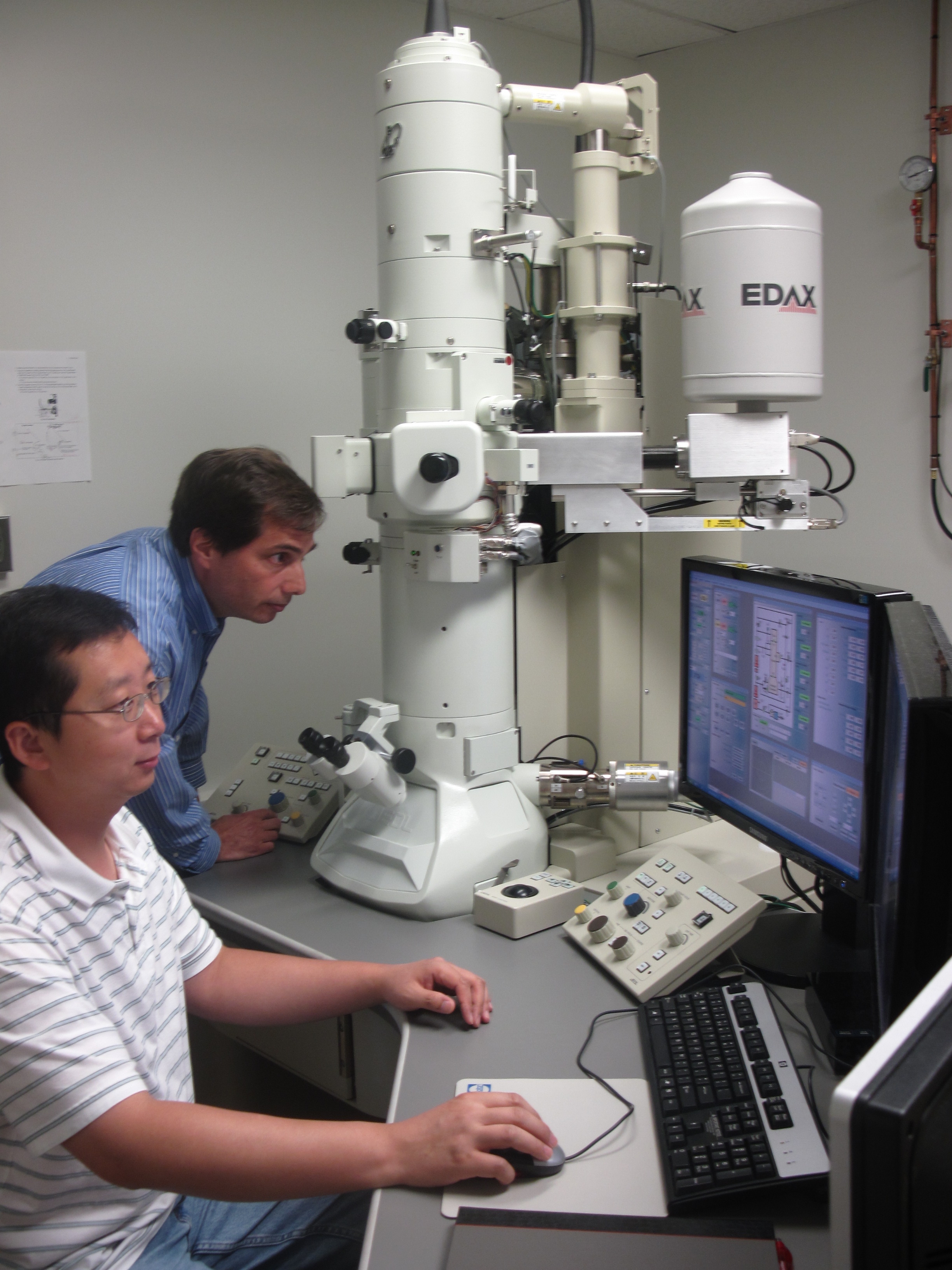
In 2009, Chemistry Professor Daniel Strongin received a $450,000 grant from the National Science Foundation to purchase a transmission electron microscope, which has a magnification capability of roughly one million, as compared to just 1,000 times for a standard optical microscope. Hutapea was one of the co-principal investigators on that grant.
“The transmission electron microscopy is a very versatile microscope and is one of the best ways to image particles or cells,” he said. “You can determine what the particle or cell looks like, its shape and size, its morphology and how its morphology changes under specific environmental conditions.”
Both Hutapea and Strongin said that up until recently, they and other Temple researchers have had to pay to substantial user fees to use scanning electron and transmission electron microscopes at other institutions, often without the necessary support to obtain sufficient images and data for their research.
Biology Professor Joel Sheffield, a microscopy expert who teaches a course in microscopy for upper level undergraduates and graduate science and engineering students, said there has been a growing trend on campus in the use of microscopy, particularly at what he calls the “super resolution advanced level,” as more and more researchers become focused on nanotechnology and nano-materials.
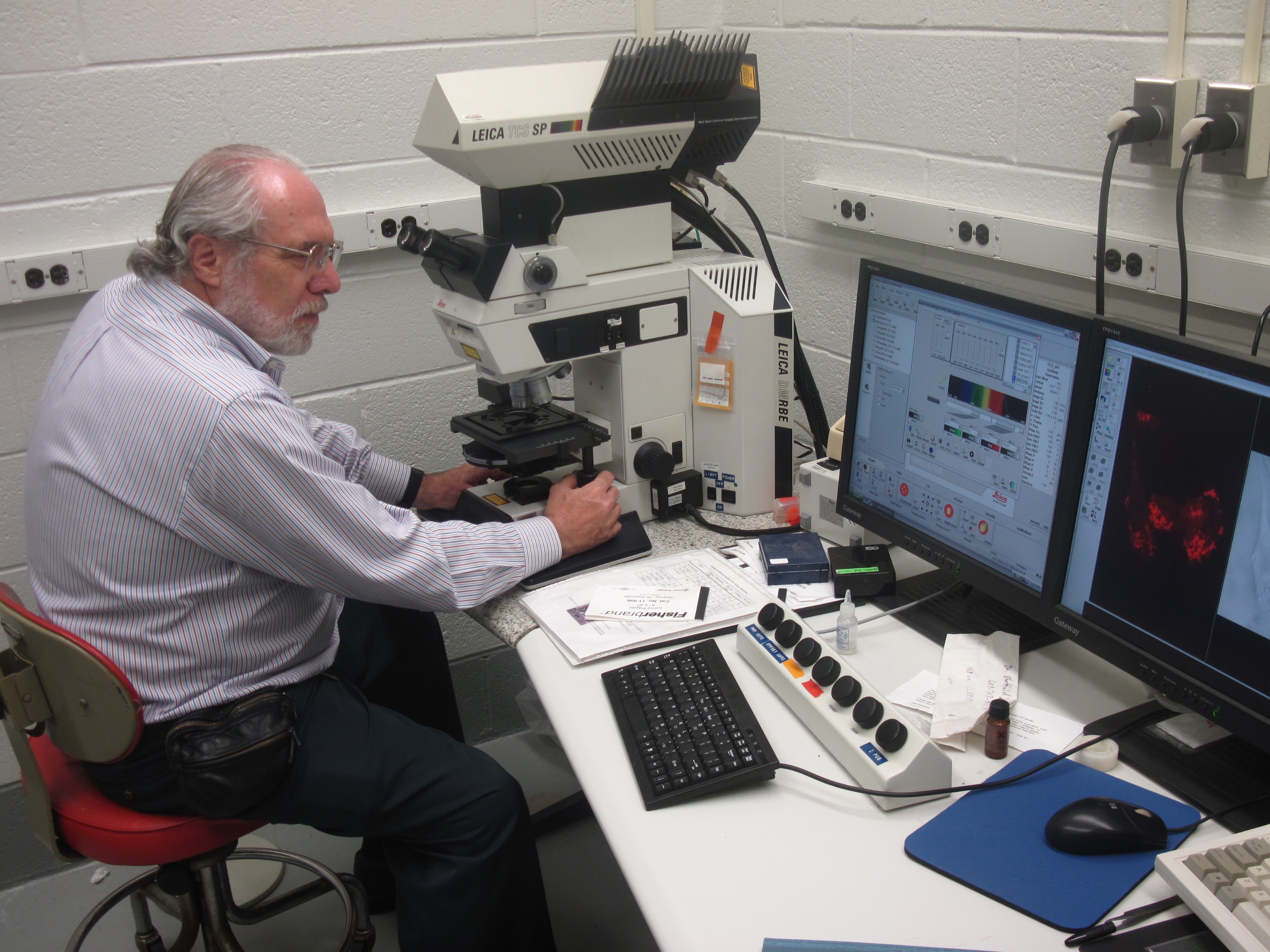
Sheffield oversees a lab facility in the Bio-Life Sciences Building that includes approximately seven different types of microscopes, including a confocal microscope, which allows researchers to reconstruct three-dimensional structures from the obtained images, and a specialized “table top field emission” scanning electron microscope purchased with $100,000 from the state Tobacco Research Funds.
Sheffield also notes that the biology department’s newest faculty member, Associate Professor Weidong Yang, has developed his own microscope for super-resolution studies of nuclear pores, the structures that allow molecules to leave or enter the cell nucleus from the cytoplasm.
“There’s been an extraordinary revolution in biology in understanding cells — looking at cellular structures and cellular processes,” said Sheffield. “At the same time, there’s been a very large growth in the field of nanotechnology. So these two things coming together have supported this movement toward enhancing microscopy capabilities at Temple through the acquisition of new microscopy instrumentation.”

